Full Length Research Paper- Asian Journal of Pharmaceutical Technology and Innovation(2024)
Radiographic study to evaluate the effect of Acetaminophen injection on orthodontic tooth movement in rats.
Maher Al Ajami1*, Feras Baba1, Abdalla Addas2 and Mohamad Yaser Abajy32Department of Histopathology, University of Aleppo, Aleppo, Syria
3Department of Biochemistry and Microbiology, University of Aleppo, Aleppo, Syria
Maher Al Ajami, Department of Orthodontics, University of Aleppo, Aleppo, Syria, Email: maheralajami328@gmail.com
Received: 11-Mar-2024, Manuscript No. AJPTI-24-129173; Editor assigned: 13-Jun-2024, Pre QC No. AJPTI-24-129173 (PQ); Reviewed: 27-Mar-2024, QC No. AJPTI-24-129173; Revised: 03-Apr-2024, Manuscript No. AJPTI-24-129173 (R); Published: 10-Apr-2024
Abstract
To evaluate the effect of acetaminophen injection as an analgesic accompanying orthodontic treatment on reducing root resorption resulting from the application of a large orthodontic force when injected during orthodontic treatment in rats. The sample consisted of 10 WISTER rats from the group of rats present in the animal house at the Faculty of Pharmacy-University of Aleppo. The rats were old for two to six months and weighed between 200-250 grams. A closing orthodontic spring made of nickel-titanium with a strength of 100 grams was applied between the upper first molar and incisor in the right and left sides of the upper jaw of the rat for a period of thirty days, and acetaminophen was injected into the right side in the gingival region closest to the upper first molar, A three-dimensional radiograph was taken before applying the orthodontic force and immediately after its removal, The distance was measured between the mesial surface of the upper second molar and the distal surface of the upper first molar in the same side. Acetaminophen injections did not have an inhibitory effect on orthodontic tooth movement like other steroidal and non-steroidal anti-inflammatory drugs.
Keywords
Acetaminophen, Tooth movement, Orthodontics, Rats, Paracetamol.
Introduction
Shortening the duration of orthodontic treatment is very important for orthodontic specialists and patients, as the negative issues related to the length of the orthodontic treatment period, such as discomfort, pain, external apical root resorption, suboptimal oral health, white spot lesions, and dental caries, become minimal [1].
Orthodontic dental treatment may be accompanied by pain for the patient, and its severity may be associated with the type of orthodontic procedure performed on the patient. Therefore, we are sometimes forced to prescribe anti-inflammatory drugs that have an analgesic effect or pain relievers to relieve this pain for the patient. Non-Steroidal Anti-Inflammatory Drugs (NSAIDs) are the most common medications in the world that. It is taken to treat signs of inflammation, including pain. Therefore, interest has increased in previous years in searching for the best pharmaceutical option for alleviating pain resulting from orthodontic treatment without affecting orthodontic tooth movement [2].
Tooth movement takes place within a diverse tissue environment that extends from fibrous ligamentous tissue to hard bone tissue [3]. The basics of dental movement must be understood to reduce the duration of treatment and achieve satisfactory results for the patient. There is much research that has dealt with the occurrence of dental movement and compared it with orthodontic forces at the molecular level. The importance of understanding the paths of this movement is that it enables us to design a better device that targets the specific cell to control it and accelerate the tooth movement [4].
Where orthodontic dental movement occurs as a result of dynamic change processes that include the shape, structure or composition of the soft and hard tissues supporting and surrounding the tooth. The dental tissues and their surroundings (dentin-cementumalveolar ligament-alveolar bone-gums) are all characterized by active restoration mechanisms and are adaptable to the mechanical forces applied to them by orthodontic devices. Orthodontic dental movement takes place as a result of mechanical stimulation, which leads to the remodeling of the alveolar bone and the Periodontal Alveolar Ligament (PDL) and when sufficient pressures are applied for a prolonged period on the tooth in the alveolar ligament area, bone resorption will occur in the direction of pressure and the tooth will move across the bone, thus the alveolar ligament plays the main role in the orthodontic movement [5].
Acetaminophen (Paracetamol)
There is a difference in the literature on whether it is considered part of the family of non-steroidal anti-inflammatory drugs or not. Its chemical composition is similar to theirs, but it lacks the antiinflammatory effect. It also lacks the blood-thinning effect and the harmful effect on the stomach lining.
This is because it inhibits the COX-3 enzyme, which it expresses. In the brain and spinal cord, it has a weak effect on COX-1 and COX-2 (and thus a weak effect on prostaglandin production) [6]. It is one of the most important and most prescribed medications in the world and is available in several pharmaceutical forms, such as capsules and tablets that are dissolved in the mouth, as a liquid given intravenously and intramuscularly, as an oral syrup, and also as rectal suppositories [7].
Experimental animals (Wister albino rats)
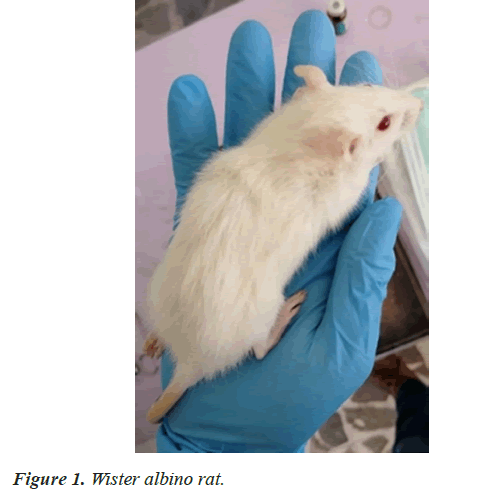
The Wister rat is albino. This breed was developed at the Wistar Institute in 1906 AD for use in biological and medical research. The Wister rat is currently one of the most common rats used in laboratory research. It is characterized by its broad head, long ears, and the length of its tail, which is always less than its body length [8,9].
They are rats belonging to the family of mammals, and their average lifespan is between two and a half years and three years. Their characteristics are cowardly, social, nocturnal, intelligent, weak eyesight, and weighing between 150-400 g. It has 16 teeth, eight in each jaw, which begin to appear on the eighth day after birth and are distributed as follows:
• Incisors: two upper and two lower. The two upper incisors are narrower, shorter, and yellower, measuring 4 mm in length and 1.5 mm in width. The two lower incisors are 7 mm long and 1.2 mm wide [10].
• Molars: Rats have six molars in each jaw, distributed into three molars on each side [11] (Figure 1).
Materials and Methods
Experimental animals
Experimental rats from the WISTER family, two to six months old when the device was applied [12], with a sample size of 10 rats taken from the animal laboratory at the Faculty of Pharmacy at the University of Aleppo, and free of systemic diseases, intra-oral diseases, and the teeth of the entire sample rats unharmed. She had not been subjected to any previous experiments or been injected with any medicinal substance.
Materials for radiological examination
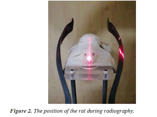
3D Cone Beam Radiography (CBCT) from the French company Carestream, the two x-rays were taken before starting the experiment and immediately after completing it while anesthetizing the rat. Ketamine was anesthetized at a dose of 87 mg/kg in the abdominal peritoneum, specifically in the triangle (button-thighgroin) [13] (Figure 2).
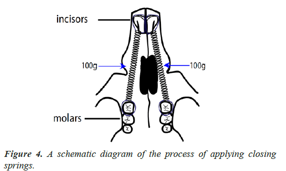
The first end of the nickel-titanium closure spring was tied with a ligature wire and tied to the upper left first molar and placed under the large circumference of the first molar and under the point of contact between the first and second upper left molar and tightened so that the ring of the closure spring was positioned in contact with the first molar and then tied the second end. From the closing spring with another ligature wire and connecting it to the upper left incisor after calibrating the strength of the spring to the force of 100 g and in the same way, the upper right first molar of the upper right incisor was bonded with a force of 100 g [14]. Then we covered the end of the ligature wire from the front end at the incisors and the back end at the first molar with a liquefied compound resin, hardening it and softening it to protect the rat’s oral mucosa and tongue from irritation. The rats were placed in numbered cages with good thermal conditions and on a soft diet. The movement was stopped after thirty days [15] (Figures 3 and 4).
Once the application of the closure springs was completed and their stability was confirmed, acetaminophen was injected at a dose of 10 mg/kg/day (AYUMI Pharmaceutical Co., Tokyo, Japan) into the right limb in the gingival region near the upper first molar in the upper jaw of the rat [16]. This dose of acetaminophen was based on the human dose and the lowest dose was used on mice in previous studies and acetaminophen was injected for three consecutive days [17].
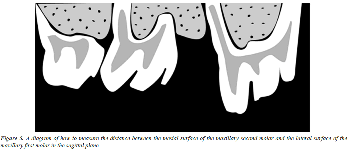
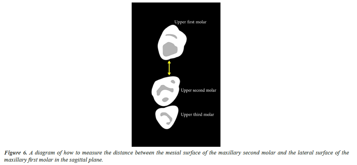
Thirty days after applying the orthodontic force, the springs were removed and the second radiograph was taken. The radiographic parameter measured the shortest distance between the lateral surface of the upper first molar and the mesial surface of the upper second molar: The distance extending from the most convex point on the mesial surface of the second molar and the most convex point on the lateral surface of the first molar on the same side was measured. This variable can be measured in the sagittal and axial sections (Figures 5 and 6).
Statistical study
Statistical analysis of the data recorded for the variable studied in the research was performed using the statistical program SPSS 20 (Statistical Package for Social Science), where the following was performed: Descriptive statistics values were calculated (number-arithmetic mean-standard deviation-smallest value- largest value-error standard) for the studied variable, and comparing the recorded values of the continuous quantitative variable with a normal distribution between the averages of the variable between the studied times to study the presence of statistically significant differences using the paired samples T-test in the statistical program SPSS 20, which is one of the parametric statistical tests that are used by for statistical analysis of continuous variables that are subject to a normal distribution in order to compare the means of the studied variable between the school periods in each group separately, the P-value of less than 0.05 was considered statistically significant (P˂0.05) at a 95% confidence level.
Dm is the shortest distance between the lateral surface of the maxillary first molar and the mesial surface of the maxillary second molar on the same side, in mm (Tables 1 and 2).
| Time | The side | Measurements | The number | SMA value | Standard deviation | Smallest value | Largest value | Standard error |
|---|---|---|---|---|---|---|---|---|
| T0 | Right | DM | 10 | 0 | 0 | 0 | 0 | 0 |
| T0 | Left | DM | 10 | 0 | 0 | 0 | 0 | 0 |
| Tf | Right | DM | 10 | 0.5 | 0.14 | 0.3 | 0.7 | 0.05 |
| Tf | Left | DM | 10 | 0.3 | 0.11 | 0.2 | 0.5 | 0.04 |
| Measurements studied | The difference between the two averages | Calculated t value | P-value | Meaning of differences |
|---|---|---|---|---|
| DM | 0.2 | 3.19 | 0.007 | There are statistically significant differences |
Results and Discussion
People undergoing orthodontic treatment often experience pain and discomfort, and many studies have classified the pain associated with orthodontic treatment as a major deterrent to orthodontic treatment [18,19]. Common medications used to relieve pain following orthodontic treatment are NSAIDs [20,21]. This effectively reduces the pain and discomfort associated with every activation of the orthodontic device, and in return, it affects orthodontic dental movement by inhibiting the inflammatory processes responsible for dental movement and bone resorption processes.
Many experiments and literature reviews have revealed that despite the great benefits of NSAIDs, they have significant side effects. Therefore, it must be given carefully, especially in children. Acetaminophen is often used as a pain reliever and fever reducer. Although it is less effective as a pain reliever than NSAIDs, it is used for all ages as a safe alternative to them due to its fewer side effects.
The experiment aims to evaluate the effect of acetaminophen injection during orthodontic treatment on the rate of orthodontic dental movement. Three-dimensional (CBCT) radiographs were taken and then an orthodontic force of 100 g was applied to the right and left sides. Acetaminophen was injected from the beginning of the experiment and the orthodontic force was applied for thirty days and then removed. Orthodontic force obtain more radiographs as soon as upon force is removal in order to assess the particular variable radiographically.
The variable denoting the shortest path between the upper first molar crown and the upper second molar crown on the same side. When comparing the averages of the shortest distance between the crowns of the upper right second molar and the crown of the upper right first molar that were injected with cetamol at the time Tf and the averages of the shortest distance between the crown of the upper left second molar and the crown of the upper left first molar that was not injected with cetamol at the time Tf, there were differences that are statistically significant. That is, when we applied a nickel-titanium orthodontic space closing spring with a force of 100 g and injected acetaminophen from the beginning of the force application on the right side, we obtained an orthodontic tooth movement greater than the orthodontic tooth movement that occurred on the left side of the upper left first molar, which received the same orthodontic force but was not injected with acetaminophen. Therefore, injecting acetaminophen during orthodontic tooth movement did not affect it and this is consistent with the study of Carmen Gonzales and his colleagues in 2009, who studied the effect of NSAIDs on orthodontic tooth movement and the resulting root resorption in rats, with a sample size of 60 rats divided into 12 groups, and by applying an orthodontic force of 50 g. Over two weeks, they found that the group that received acetaminophen had greater orthodontic tooth movement compared to the groups that received doses of NSAIDs. The study of Masato Kaku and his colleagues in 2019, who studied the evaluation of the effect of acetaminophen on orthodontic tooth movement and the resulting root resorption by applying an orthodontic force of 10 g over thirty days, also agreed. They found that there were no statistically significant differences between the group of rats that were injected with acetaminophen and the group. Who were not injected with acetaminophen, i.e. acetaminophen did not have an inhibitory effect on orthodontic tooth movement. We agreed with the study of Juhn J. Roche and his colleagues in 1997 AD, who studied the effect of acetaminophen on orthodontic dental movement in rabbits, and by radiological evaluation they found that it does not affect orthodontic dental movement and is the best analgesic for people receiving orthodontic treatment. We agreed with the study of A.C. Stabile and his colleagues in 1997, who studied the effect of acetaminophen and celecoxib on orthodontic tooth movement and concluded that they do not affect orthodontic tooth movement. We disagreed with the study of Shaza M. Hammad and her colleagues in 2012, who studied the effect of several analgesics on orthodontic tooth movement in rats and found that acetaminophen interferes with orthodontic tooth movement. The reason for this is that she gave a large dose of acetaminophen of 150 mg/kg daily for two months, and this indicates the long duration of drug administration and its effectiveness in the rat’s body.
Conclusion
Injection of acetaminophen into the area near the maxillary first molar during orthodontic treatment did not inhibit orthodontic tooth movement and accelerated orthodontic tooth movement compared to the side not injected with acetaminophen.
These results are consistent with previous studies highlighting the efficacy of acetaminophen in pain management during orthodontic treatment. The growing body of evidence supporting its use as a safe and effective analgesic for orthodontic patients. By providing clinicians with additional therapeutic options, efforts to minimize the duration of orthodontic treatment can be enhanced, ultimately improving patient satisfaction and oral health outcomes.
References
- Rashid A, ElSharaby FA, Nassef EM, et al. Effect of platelet‐rich plasma on orthodontic tooth movement in dogs. Orthod Craniofac Res. 2017; 20(2):102-10.
[Crossref][Google Scholar] [Pubmed]
- Gonzales C, Hotokezaka H, Matsuo KI, et al. Effects of steroidal and nonsteroidal drugs on tooth movement and root resorption in the rat molar. Angle Orthod. 2009; 79(4):715-26.
[Crossref][Google Scholar] [Pubmed]
- Nanda R. Biomechanics and esthetic strategies in clinical orthodontics. Elsevier Health Sciences; 2005.
- Isola G, Matarese G, Cordasco G, et al. Mechanobiology of the tooth movement during the orthodontic treatment: a literature review. Minerva stomatol. 2016.
[Google Scholar] [Pubmed]
- Proffit WR, Fields H, Larson B, et al. Contemporary orthodontics: Contemporary orthodontics-e-book. Elsevier Health Sciences; 2018.
- Bartzela T, Türp JC, Motschall E, et al. Medication effects on the rate of orthodontic tooth movement: a systematic literature review. Am J Orthod Dentofacial Orthop. 2009;135 (1):16-26.
[Crossref][Google Scholar] [Pubmed]
- Saccomano SJ. Acute acetaminophen toxicity in adults. Nurse Pract. 2019; 44(11):42-47.
[Crossref][Google Scholar] [Pubmed]
- Clause BT. The wistar institute archives: rats (not mice) and history. Mendel Newsl. 1998;7(2).
[Google Scholar] [Pubmed]
- Clause BT. The wistar rat as a right choice: Establishing mammalian standards and the ideal of a standardized mammal. J Hist Biol. 1993;26(2):329-49.
[Crossref][Google Scholar] [Pubmed]
- Hauber GG, Nouer DF, Pereira NJS, et al. Effects of short-and long-term celecoxib on orthodontic tooth movement. Angle Orthod. 2008; 78(5):860-5.
[Crossref][Google Scholar] [Pubmed]
- Ino KA, Hotokezaka H, Kondo T, et al. Lithium chloride reduces orthodontically induced root resorption and affects tooth root movement in rats. Angle Orthod. 2018; 88(4):474-82.
[Crossref][Google Scholar] [Pubmed]
- Nakano T, Hotokezaka H, Hashimoto M, et al. Effects of different types of tooth movement and force magnitudes on the amount of tooth movement and root resorption in rats. Angle Orthod. 2014; 84(6):1079-85.
[Crossref][Google Scholar] [Pubmed]
- Kaku M, Yamamoto T, Yashima Y, et al. Acetaminophen reduces apical root resorption during orthodontic tooth movement in rats. Arch Oral Biol. 2019; 102:83-92.
[Crossref][Google Scholar] [Pubmed]
- Carlos F, Cobo J, Perillan C, et al. Orthodontic tooth movement after different coxib therapies. Eur J Orthod. 2007; 29(6):596-9.
[Crossref][Google Scholar] [Pubmed]
- Haynes S. Discontinuation of orthodontic treatment relative to patient age. J Dentistry. 1974; 2(4):138-42.
[Crossref][Google Scholar] [Pubmed]
- Kluemper GT, Hiser DG, Rayens MK, et al. Efficacy of a wax containing benzocaine in the relief of oral mucosal pain caused by orthodontic appliances. Am J Orthod Dentofacial Orthop. 2002 ;122(4):359-65.
[Crossref][Google Scholar] [Pubmed]
- Krishnan V, Davidovitch Z. The effect of drugs on orthodontic tooth movement. Orthodon Craniofac Res. 2006;9(4):163-71.
[Crossref][Google Scholar] [Pubmed]
- Krishnan V. Orthodontic pain: from causes to management—a review. Eur J Orthod. 2007; 29(2):170-9.
[Crossref][Google Scholar] [Pubmed]
- Hauber GG, Nouer DF, Pereira NS, et al. Effects of short-and long-term celecoxib on orthodontic tooth movement. Angle Orthod. 2008; 78(5):860-5.
[Crossref][Google Scholar] [Pubmed]
- Zhang W, Nuki G, Moskowitz RW, et al.OARSI recommendations for the management of hip and knee osteoarthritis: part III: Changes in evidence following systematic cumulative update of research published through January 2009. Osteoarthritis Cartilage. 2010; 18(4):476-99.
[Crossref][Google Scholar] [Pubmed]
- Zhang W, Jones A, Doherty M. Does paracetamol (acetaminophen) reduce the pain of osteoarthritis?: a meta-analysis of randomised controlled trials. Ann Rheum Dis. 2004; 63(8):901-7.
[Crossref][Google Scholar] [Pubmed]
Citation: Ajami MA, Baba F, Addas A, et al. Radiographic study to evaluate the effect of Acetaminophen injection on orthodontic tooth movement in rats. AJPTI 2024;12 (46):1-5.

